Smooth Muscle Diagram : Smooth Muscle Cells In Vascular Remodeling After Injury. It is found in the walls of ducts and blood and lymphatic vessels, as well as in the walls of the digestive, respiratory and urogenital tracts. Smooth muscle cells are found in the dividers of empty organs, including the stomach, digestion tracts, urinary bladder and uterus, and in the dividers of paths, for example, the supply routes and veins of the circulatory framework, and the tracts of the. The cells are spindle shaped, and the nucleus is central. 12 photos of the smooth muscle diagram. Smooth muscle tissue, unlike striated muscle, contracts slowly and automatically.
It is divided into two subgroups; Vascular smooth muscle refers to the particular type of smooth muscle found within, and composing the majority of the wall of blood vessels. It is the pen diagram of skeletal, smooth and cardiac muscle for class 10, 11 and 12. The muscle tissue (patrick steele) Smooth muscle, muscle that shows no cross stripes under microscopic magnification.
Human Smooth Muscle How Many Smooth Muscles In The Human Body Smooth Muscle Tissue Skeletal Muscle Muscle Anatomy Muscular System from i.pinimg.com By ning zhou, shaunrick stoll. In arteries, smooth muscle movements maintain the arteries' diameter. It is divided into two subgroups; Friends in this video i will tell you about how to draw structure of smooth muscle fibre. Vascular smooth muscle is innervated primarily by the sympathetic nervous system through adrenergic receptors (adrenoceptors). It is the pen diagram of skeletal, smooth and cardiac muscle for class 10, 11 and 12. Smooth muscle is made up of cells that contain a single central nucleus. The pupillary sphincter muscle in your eye is a smooth muscle that shrinks the size of your.
In arteries, smooth muscle movements maintain the arteries' diameter.
In arteries, smooth muscle movements maintain the arteries' diameter. Smooth muscle tissue, unlike striated muscle, contracts slowly and automatically. Smooth muscle determines the flow of blood in the arteries. Smooth muscles have a much stronger ability to contract than skeletal. The three types present are: The cells stick together and are connected by specialised cell junctions, called gap junctions. Smooth muscles are unique in their largely involuntary response, and in their structure. It also occurs in the spleen (capsule and trabeculae), eye (iris and ciliary body), skin (arrector pili muscles of hairs. They average a length of about 0.2 mm. Smooth muscles in a woman's uterus (or womb) help to push babies out of the body during childbirth. It is found in the walls of ducts and blood and lymphatic vessels, as well as in the walls of the digestive, respiratory and urogenital tracts. Smooth muscle contracts under certain stimuli as atp is freed. Smooth muscles in arteries and veins are largely responsible for regulation of blood pressure.
Smooth muscle anatomy smooth muscle tissue is also known as visceral muscle tissue. The muscle tissue (patrick steele) The cells are spindle shaped, and the nucleus is central. Smooth muscle (textus muscularis levis) smooth muscle is a type of tissue found in the walls of hollow organs, such as the intestines, uterus and stomach. The cells stick together and are connected by specialised cell junctions, called gap junctions.
Types Of Smooth Muscle Medical Physiology Euroform Healthcare from www.euroformhealthcare.biz • smooth muscles respond to stretch only briefly, and then adapts to its new length • the new length however, retains its original _____ seconds or minutes after it has been elongated or shortened (e.g. Smooth muscles have a much stronger ability to contract than skeletal. Smooth muscle anatomy smooth muscle tissue is also known as visceral muscle tissue. It is found in the walls of ducts and blood and lymphatic vessels, as well as in the walls of the digestive, respiratory and urogenital tracts. Smooth muscle cells are found in the dividers of empty organs, including the stomach, digestion tracts, urinary bladder and uterus, and in the dividers of paths, for example, the supply routes and veins of the circulatory framework, and the tracts of the. Smooth muscle tissue, unlike striated muscle, contracts slowly and automatically. Thousands, or even tens of thousands, of small fibers make up each muscle. This smooth muscle can be found surrounding the walls of the blood vessels, the bronchioles in the lungs, and the sphincter muscles used in the gi tract.the gi tract, which is tubular by design, also houses longitudinal muscles in addition to the smooth.
Smooth muscles have a much stronger ability to contract than skeletal.
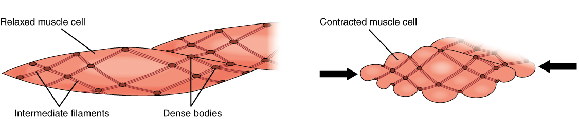
In this video i have shown the simplest way of drawing muscle drawing. 12 photos of the smooth muscle diagram. In addition, the contractile state of smooth muscle is controlled by hormones, autocrine/paracrine agents, and other local chemical signals. Smooth muscle histology and diagram (inlet). Smooth muscle (textus muscularis levis) smooth muscle is a type of tissue found in the walls of hollow organs, such as the intestines, uterus and stomach. Diagram of smooth muscle contraction, smooth cardiac and skeletal muscle diagram, smooth muscle cell diagram, smooth muscle cell picture, smooth muscle contraction diagram, human muscles, diagram of smooth muscle contraction, smooth cardiac and skeletal muscle diagram, smooth muscle cell diagram, smooth. The smooth muscles perform the functions in the contrast of other types of muscles. Smooth muscles exhibits a phenomenon called _____ in which: After watching this video completely you will draw this diagram very easily. Smooth muscle has a fusiform shape, which resembles a football or spindle. Smooth muscle is made up of cells that contain a single central nucleus. It is the pen diagram of skeletal, smooth and cardiac muscle for class 10, 11 and 12. 12 photos of the smooth muscle diagram.
Stomach tissues types and structure infographic diagram including smooth muscle loose connective nervous blood. Arteries have thick walls due to smooth muscle cells, which help them carry blood away from the heart to every part of. Diagram of smooth muscle contraction, smooth cardiac and skeletal muscle diagram, smooth muscle cell diagram, smooth muscle cell picture, smooth muscle contraction diagram, human muscles, diagram of smooth muscle contraction, smooth cardiac and skeletal muscle diagram, smooth muscle cell diagram, smooth. Smooth muscles in arteries and veins are largely responsible for regulation of blood pressure. It constitutes much of the musculature of
Smooth Muscle Anatomy And Physiology from opentextbc.ca It is the pen diagram of skeletal, smooth and cardiac muscle for class 10, 11 and 12. By ning zhou, shaunrick stoll. The pupillary sphincter muscle in your eye is a smooth muscle that shrinks the size of your. Asapknowledge posted a video to playlist educational videos. Smooth muscles are unique in their largely involuntary response, and in their structure. 12 photos of the smooth muscle diagram. Smooth muscles exhibits a phenomenon called _____ in which: The three types present are:
Vascular smooth muscle is innervated primarily by the sympathetic nervous system through adrenergic receptors (adrenoceptors).
This diagram shows a few of the cells that can be seen in the stained section below. Smooth muscle is widely distributed in the body. Vascular smooth muscle refers to the particular type of smooth muscle found within, and composing the majority of the wall of blood vessels. Smooth muscle determines the flow of blood in the arteries. In skeletal muscle, a single type of somatic nervous system traverses to muscle, where it stimulates organelle in the muscle cells in order to release calcium. It is layered in a distinctive pattern of circular layers. Smooth muscle contracts under certain stimuli as atp is freed. After watching this video completely you will draw this diagram very easily. Smooth muscles are unique in their largely involuntary response, and in their structure. Smooth muscle anatomy smooth muscle tissue is also known as visceral muscle tissue. Vascular smooth muscle is innervated primarily by the sympathetic nervous system through adrenergic receptors (adrenoceptors). In this video i have shown the simplest way of drawing muscle drawing. Smooth muscle (textus muscularis levis) smooth muscle is a type of tissue found in the walls of hollow organs, such as the intestines, uterus and stomach.